EEG Variants: Routine EEG, Ambulatory EEG and QEEG Testing
Integrative Care for Concussion / Head Injury Sufferers
in Ft. Lauderdale, Tampa, and Orlando, Florida
Brain Wave Diagnostics: Understanding the Significance of EEG Testing
An Electroencephalogram (EEG) test is a non-invasive neurodiagnostic technique that records the electrical activity of the brain. An Electroencephalogram (EEG) test is used to evaluate several types of brain disorders.
Why would a doctor order an EEG test?
A doctor might order an EEG (electroencephalogram) test for several reasons:
Diagnosis of Epilepsy and Seizure Disorders: EEG is commonly used to diagnose and monitor epilepsy and other seizure disorders. It helps in identifying abnormal electrical activity in the brain that is characteristic of seizures.
Evaluation of Brain Disorders: EEG can be used to diagnose other brain disorders such as tumors, infections, encephalitis, and encephalopathy.
Assessment of Brain Damage: It can help assess the extent of brain damage following head injuries or strokes.
Monitoring during Surgery: EEG might be used to monitor brain activity during certain types of surgeries to ensure the brain is functioning properly.
Sleep Disorders: It can be used to diagnose sleep disorders like narcolepsy or sleep apnea by monitoring brain activity during sleep.
Unexplained Coma: EEG can help determine the level of brain activity in patients who are in a coma.
What can an EEG show that an MRI cannot?
An EEG (electroencephalogram) and an MRI (magnetic resonance imaging) provide different types of information about the brain, and each has unique advantages.
EEG:
- Real-Time Brain Activity: An EEG measures electrical activity in the brain, providing real-time data on brain function. This is particularly useful for diagnosing conditions like epilepsy, where abnormal electrical activity needs to be detected as it occurs.
- Seizure Detection: EEGs are more effective than MRIs for detecting seizures, which often do not show structural abnormalities on an MRI. EEGs can capture the rapid and transient electrical disturbances that characterize seizures.
- Functional Insights: EEGs are useful for assessing brain activity during various states, such as wakefulness, sleep, and cognitive tasks. This makes them valuable for diagnosing sleep disorders, monitoring anesthesia depth, and investigating cognitive and attention disorders like ADHD.
MRI:
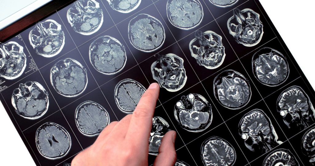
- Structural Imaging: MRIs provide detailed images of brain structures, allowing for the detection of physical abnormalities such as tumors, lesions, and brain injuries. This makes MRI the preferred tool for identifying structural issues within the brain.
- No Real-Time Data: Unlike EEGs, MRIs do not provide information on brain activity in real time. They are static images that capture the brain’s structure at a single point in time.
In summary, while an EEG is superior for detecting and monitoring electrical activity and functional brain abnormalities, an MRI excels in providing detailed anatomical images. The choice between the two depends on the specific clinical question and condition being investigated.
Routine EEG Test
Routine EEG is a diagnostic test that involves recording brain wave patterns over a short period, typically lasting from 20 to 60 minutes. The procedure is conducted in a clinical setting and is often used to detect abnormalities such as epileptic seizures, brain injuries, and certain sleep disorders.
Key features of EEG Test:
- Test Duration: 20 to 60 minutes
- Appointment Duration: 1-2 hours
- Setting: EEG is typically conducted in a quiet room to minimize external interference with the recording.
- Purpose: Detecting and diagnosing neurological conditions, including epilepsy and sleep disorders. The EEG may also be used to determine the overall electrical activity of the brain to evaluate trauma.
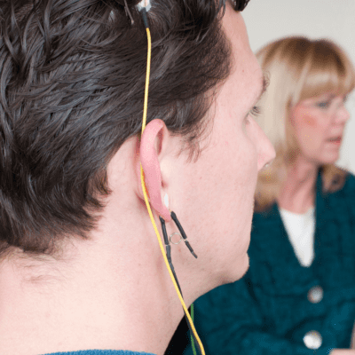
- Procedure: Electrodes are attached to the scalp to record brain wave patterns. The procedure may vary depending on your condition.
Patient Preparation: It’s important for the patient to be relaxed and comfortable during the test, so they may be asked to avoid caffeine or alter their medication schedule as advised by their healthcare provider.
- Recording Brain Waves: The EEG machine records the electrical activity produced by the brain in the form of waves. This recording is typically done while the patient is at rest with their eyes closed and then repeated with their eyes open.
Monitoring: An EEG technologist or EEG technician monitors the recording during the procedure to ensure the quality of the data and to identify any issues that may need to be addressed.
Analysis: A neurologist or healthcare provider analyzes the recorded EEG data. They interpret the patterns of brain waves and look for abnormalities that may indicate neurological conditions such as epilepsy, sleep disorders, or other brain disorders.
To learn more about EEG, Click Here
QEEG Test
QEEG, also known as brain mapping, is an advanced form of EEG that involves quantitatively analyzing electrical activity in the brain. Unlike Routine EEG, QEEG provides detailed insights into brain function by analyzing brain waves’ frequency, amplitude, and coherence. QEEG is commonly used to assess cognitive function, identify neurological disorders, and guide neurofeedback interventions.
Key features of the QEEG Test:
- Test Duration: 20 minutes
- Appointment Duration: 60 minutes
- Setting: Clinical environment.
- Purpose: Quantitative analysis of brain wave patterns to assess cognitive function and diagnose neurological disorders.
- Procedure: Electrodes are attached to the scalp to record brain wave patterns.
Patient Preparation: The patient needs to be adequately prepared for the QEEG procedure. This may include providing information about any medications they are taking and ensuring they are in a relaxed state.
- Recording Brain Waves: The QEEG system records the brain’s electrical activity over a specified period, typically while the patient is at rest with their eyes closed and open. This recording captures baseline brain activity and allows for the analysis of various brain wave patterns.
Monitoring: An EEG technologist or EEG technician monitors the recording during the procedure to ensure the quality of the data and to identify any issues that may need to be addressed.
Analysis: A neurologist or healthcare provider analyzes the recorded EEG data. They interpret the patterns of brain waves and look for abnormalities that may indicate neurological conditions such as epilepsy, sleep disorders, or other brain disorders.
Data Analysis: The recorded data is analyzed quantitatively using specialized software. This analysis involves examining the frequency, amplitude, and coherence of the recorded brain waves.
Mapping Brain Activity: The software generates maps and graphs that represent the distribution of brain activity across different regions. These maps provide a visual representation of the quantitative information obtained from the analysis.
Comparison to Normative Databases: The results of the QEEG are often compared to normative databases that contain data from healthy individuals of similar age and gender. This step helps identify patterns or deviations that may indicate abnormalities.
Report Generation: A comprehensive report is generated based on the analysis. This report typically includes information about brain wave patterns, any deviations from the norm, and their potential implications. It may also provide recommendations for further evaluation or intervention.
Clinical Interpretation: A trained healthcare professional, often a neurologist or a neurofeedback practitioner, interprets the results in a clinical context. The interpretation considers the patient’s specific symptoms, medical history, and any other relevant information.
- Diagnostic and Treatment Planning: The findings from the QEEG may contribute to the diagnosis of neurological conditions or inform treatment planning. In some cases, QEEG results are used to guide neurofeedback interventions, where individuals can learn to self-regulate their brain activity.
To learn more about QEEG, Click Here
Ambulatory EEG (24 hrs/48 hrs/72 hrs)
Ambulatory EEG, also known as long-term video EEG monitoring, extends the recording duration to 24, 48, or 72 hours. This type of EEG is crucial for capturing intermittent or rare neurological events, such as seizures, that may not be evident during a short-duration recording. Ambulatory EEG is particularly valuable in epilepsy monitoring and evaluating patients with suspected but infrequent neurological events.
Key features of the Ambulatory EEG Test:
- Duration: Extended (24 to 72 hours).
- Setting: Can be conducted in a hospital or at home.
- Purpose: Capturing intermittent neurological events, particularly in epilepsy monitoring.
- Procedure: Electrodes are attached to the scalp to record brain wave patterns using portable recording equipment worn by the patient during their daily activities. The device is usually worn in a small pouch or belt around the waist to ensure it is discreet and doesn’t interfere with the patient’s routine.
Understanding QEEG Brain Mapping and its Interpretation by BCN Therapists
Quantitative Electroencephalography (QEEG) brain mapping is a sophisticated neuroimaging technique used to assess the electrical activity of the brain. This method involves the use of electroencephalography (EEG) to record brain wave patterns, which are then analyzed to produce a detailed map of brain function. The interpretation of these maps is crucial for diagnosing and treating various neurological and psychological conditions.
This post explores who is qualified to interpret QEEG brain maps and delves into the role of Board Certified Neurofeedback (BCN) Therapists at Radius TBI in the EEG recording and QEEG analysis process. To learn more, please visit who interprets a QEEG brain map.
Routine EEG, QEEG, and Ambulatory EEG
are distinct forms of electroencephalography, each serving unique purposes in the diagnosis and monitoring of neurological conditions. Routine EEG provides a snapshot of brain activity, while QEEG adds a quantitative dimension for in-depth analysis. Ambulatory EEG, with its extended recording duration, is invaluable in capturing intermittent neurological events, offering a comprehensive view of brain function over an extended period. The combined use of these EEG variants enhances the ability of healthcare professionals to diagnose, monitor, and tailor treatment plans for patients with various neurological disorders.
Have you ever experienced brain fog, difficulty concentrating, or other cognitive issues? If so, you may benefit from a QEEG test. A non-invasive way of measuring brain activity. Contact us now!
DID YOU KNOW?
TBI is a major cause of death and life-long disability in the United States. (including all levels of severity)
- An estimated 1.5 million Americans sustain a TBI (Sosin, Sniezek and Thurman 1996);
- 50,000 die from these injuries; and 80,000 to 90,000 experience onset of long-term disability (CDC 1999).
- An estimated 5.3 million Americans live with a permanent TBI-related disability today (CDC 1999).
According to the report to Congress on mild traumatic brain injury in the United States. Centers for Disease Control and Prevention; 2003. https://www.cdc.gov/traumaticbraininjury
Blogs

Meet Our No.1 Best Neuropsychologist in Tampa, FL Location
At Radius TBI, we pride ourselves on providing exceptional care for individuals suffering from traumatic brain injuries (TBI) and concussions. Our integrated medical team in Tampa, FL, includes some of the most respected and experienced professionals in the field, ensuring
VNG Testing: A Vital Tool in Evaluating Vertigo and Dizziness Disorders
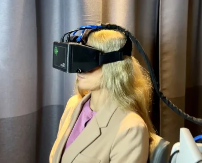
VNG testing can be important in diagnosing and evaluating balance issues after a traumatic brain injury. This test is non-invasive, safe, and effective at evaluating the inner ear, eye movements, and nervous system. If you’re experiencing balance issues after a

Workers Compensation Neurologist in Florida
We Are Dedicated TBI Team & We Are Accepting Workers Compensation. Call Us at 954-900-5072. We Are Here For Your Brain Injury Recovery.
Concussion from A Car Accident Hidden Dangers You Must Know
Take control of your brain health with effective Neurofeedback Therapy Service at our medical centers in Florida. Get help now! Call 954-900-5072
Neurofeedback Therapy: The Brain Treatment For Traumatic Brain Injury and Concussion Patients
Take control of your brain health with effective Neurofeedback Therapy Service at our medical centers in Florida. Get help now! Call 954-900-5072
Meet Skilled One Of The Top Neurologist In Tampa: Radius TBI
Have you injured your head? Call 813-736-1444 to get checked by our one of the top neurologist in Tampa. Transforming your lives through neurology

Your Premier TBI/Concussion Center in Tampa, FL
At Radius TBI, our centers in Ft. Lauderdale, Tampa, and Orlando, Florida, we are dedicated to providing exceptional care tailored to individuals with head injuries. This blog will explore why choosing Radius TBI as your trusted TBI/Concussion Center is the

Why See Neuropsychologist After Head Injury?
A head injury can be a devastating event that can have long-lasting effects on a person’s life. Depending on the severity of the injury, a person may experience a range of physical, cognitive, and emotional symptoms that can significantly impact


