Are you struggling with dizziness, vertigo, or unexplained balance issues?
Discover the non-invasive VNG testing for dizziness, vertigo, and balance disorders. Find the root cause and regain control.
Talk to our medical team of skilled healthcare professionals about VNG testing if you frequently experience dizziness, vertigo, or balance problems. Finding the root of your symptoms will enable you to improve your overall well-being and live a better life.
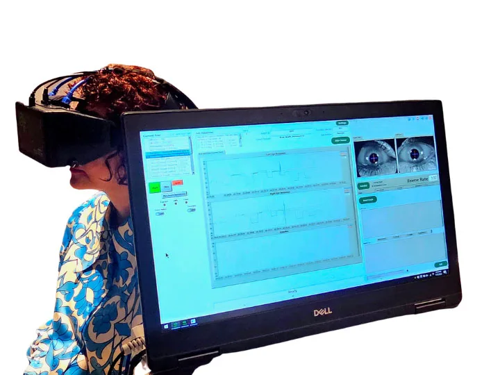
You may benefit from a diagnostic test called videonystagmography (VNG). VNG is a non-invasive assessment healthcare professionals use to identify the root cause of balance disorders and determine the most effective treatment.
Understanding Videonystagmography (VNG): The Key to Diagnosing Balance Disorders
Videonystagmography (VNG) is a critical diagnostic tool used to evaluate patients experiencing dizziness, vertigo, or balance disorders. It provides an in-depth look at the inner ear and the brain’s ability to coordinate eye movements and maintain balance. If you’re considering VNG testing or have recently undergone it, understanding what a normal result means is essential for your health journey.
How Does VNG Work?
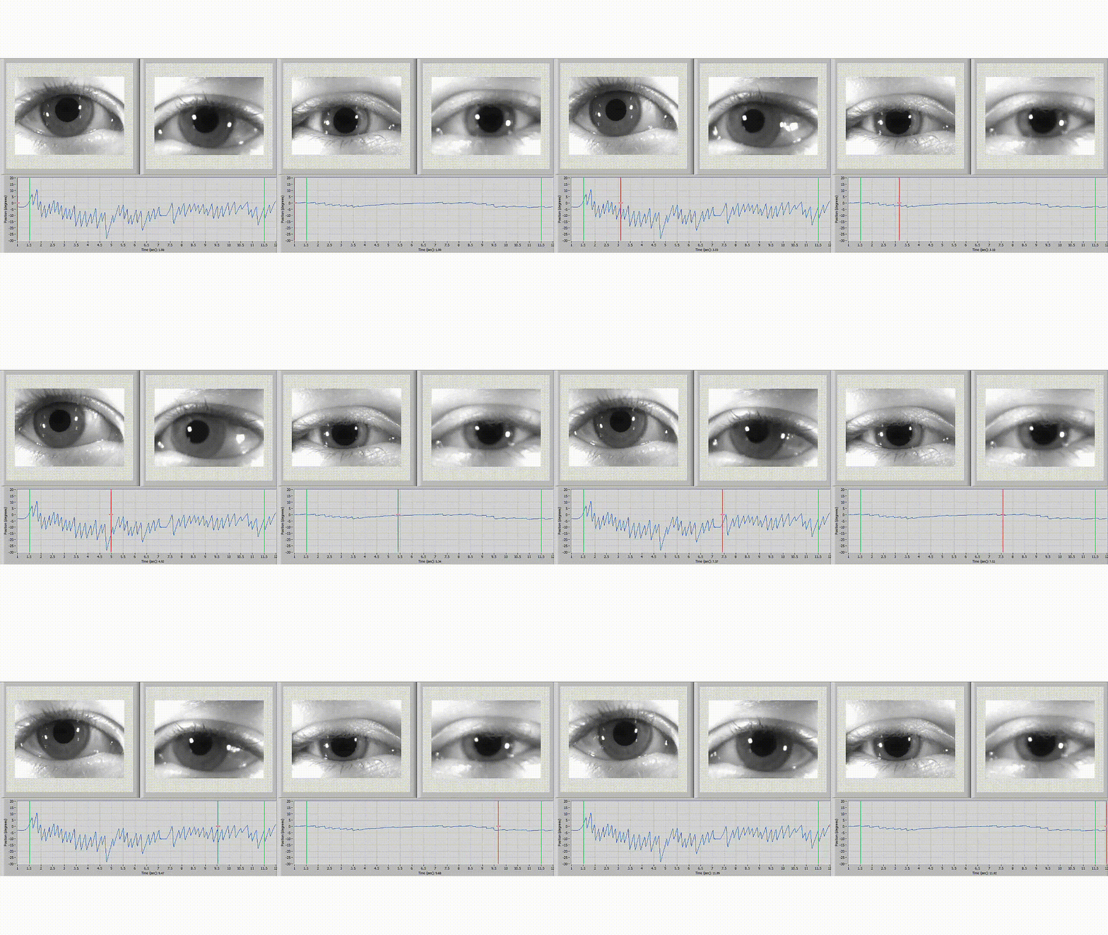
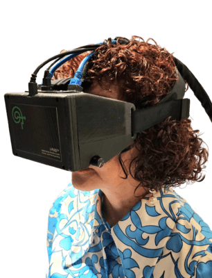
A VNG test participant will put on specialized goggles with cameras to track their eye movements in the infrared spectrum. Due to the close relationship between the vestibular system and the eyes, eye movement analysis is a reliable technique to assess inner ear function.
The VNG test May consists of two to Four main components:
A VNG test participant will put on specialized goggles with cameras to track their eye movements in the infrared spectrum. Due to the close relationship between the vestibular system and the eyes, eye movement analysis is a reliable technique to assess inner ear function.
- Oculomotor tests: Evaluate the patient’s eye movements as they track an object in the visual field, such as a dot on a screen. This aids in assessing the central nervous system’s functionality and capacity to regulate eye movements.
- Optokinetic Nystagmus: The patient tracks a series of moving stripes to evaluate eye tracking and reflexive eye movements.
- Positional tests: The head and body of the patient will be moved into different positions to gauge how the vestibular system reacts to these movements. This can help find problems with the sensory organs in the inner ear or the nerves that link them to the brain.
- Caloric Testing: Each ear is irrigated with warm and cool air or water to stimulate the inner ear and observe the resulting eye movements.
Interpreting VNG Results
Once the VNG tests are completed, the results can either be normal or abnormal.
Normal VNG Results
A normal VNG result indicates that the inner ear and the brain’s balance processing systems are functioning correctly. This means:
- No Significant Abnormalities: The patient’s eye movements are within the normal range, and there are no signs of vestibular dysfunction.
- Further Investigation Needed: If the patient continues to experience symptoms despite normal VNG results, other causes outside the vestibular system may need to be explored. This could include neurological conditions, cardiovascular issues, or psychological factors.
Abnormal VNG Results
An abnormal VNG result points to potential issues within the vestibular system. These results can help pinpoint the exact nature of the problem:
- Peripheral Vestibular Disorders: Conditions like benign paroxysmal positional vertigo (BPPV), vestibular neuritis, or Ménière’s disease may be indicated by abnormal VNG results.
- Central Vestibular Disorders: Abnormalities might also suggest issues in the brainstem or cerebellum, such as multiple sclerosis or stroke.
- Customized Treatment Plan: Based on the specific findings, a targeted treatment plan will be developed. This could include vestibular rehabilitation therapy, medication, lifestyle modifications, or in some cases, surgical interventions.
Next Steps After VNG Testing
- Consultation with Healthcare Provider: Discuss the VNG results with your healthcare provider to understand the implications fully.
- Develop a Treatment Plan: If abnormalities are detected, your provider will create a treatment plan tailored to address the specific vestibular disorder.
- Follow-Up Tests: Additional testing or referrals to specialists (e.g., neurologists, otolaryngologists) may be necessary for comprehensive care.
- Lifestyle and Home Adjustments: Depending on the diagnosis, making changes at home and in daily activities to manage symptoms and improve quality of life may be recommended.
VNG testing is a valuable diagnostic tool for identifying the underlying causes of dizziness and balance disorders. Whether the results are normal or abnormal, understanding what they mean and the subsequent steps can significantly impact the management and treatment of these conditions. Always consult with your healthcare provider to ensure you receive the most appropriate care based on your VNG results.
Who Can Benefit from VNG Testing?
Individuals experiencing unexplained dizziness, vertigo, or balance issues may benefit from VNG testing. Some common vestibular disorders that can be diagnosed with VNG include:
- Concussion/TBI
- Benign paroxysmal positional vertigo (BPPV)
- Meniere’s disease
- Vestibular neuritis or labyrinthitis
- Vestibular migraine
Additionally, VNG test can aid in diagnosing central nervous system conditions like traumatic brain injury, multiple sclerosis, or cerebral ataxia that can manifest as vertigo or balance problems.
Videonystagmography (VNG) is an essential diagnostic tool for identifying the root cause of balance disorders and guiding effective treatment plans.
One of the benefits of VNG test diagnose is that it is non-invasive and relatively quick. The test can usually be completed within 30 to 60 minutes, and there is no need for sedation or anesthesia.
Additionally, VNG testing is highly sensitive and can detect even subtle abnormalities in the vestibular system, making it an essential tool for diagnosing and managing balance disorders.
DID YOU KNOW?
One study found that a large percentage of people with traumatic brain injury (90%) or a stroke (86.7%) had issues with their eye movements. Specifically, problems with focusing and aligning the eyes were more common in those with traumatic brain injury, while crossed eyes and problems with a nerve called the cranial nerve were more common in those who had a stroke. The difficulty in moving the eyes in a coordinated way was similar in both groups.
Reference: Ciuffreda KJ, Kapoor N, Rutner D, et al. Occurrence of oculomotor dysfunctions in acquired brain injury: a retrospective analysis. Optometry. 2007;78:155-161
Read more: pubmed.ncbi.nlm.nih.gov
Latest News & Updates
Blogs
Recent Blogs
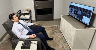
In the world of neuroscience, one of the advanced tools used to understand brain function is the Quantitative Electroencephalogram (QEEG), often referred to as brain mapping. But who interprets these intricate brain maps, and why

Meet Our No.1 Best Neuropsychologist in Tampa, FL Location
At Radius TBI, we pride ourselves on providing exceptional care for individuals suffering from traumatic brain injuries (TBI) and concussions. Our integrated medical team in Tampa, FL, includes some of the most respected and experienced
Did you know? Dealing with a traumatic brain injury (TBI) often means navigating a range of complications, one of the most prevalent being sleep disturbances. Surprisingly, 30-70% of individuals with a TBI experience some form
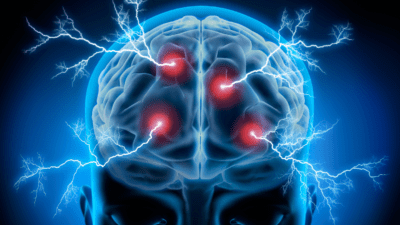
Understanding Routine EEG, QEEG, and Ambulatory EEG Tests
EEG, QEEG, and Ambulatory EEG are distinct forms of electroencephalography, each serving unique purposes in the diagnosis and monitoring of neurological conditions.


