Maximizing TBI Recovery: The Benefits of DTI Testing for Treatment Planning
Integrative Care for Concussion / Traumatic Brain Injury Sufferers
in Broward County & Hillsborough County
DTI imaging is a valuable tool for diagnosing and managing TBI-related injuries. It is a non-invasive, highly sensitive test that can provide important information about the structural integrity of the brain’s white matter.
What is DTI Testing?
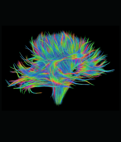
Diffusion Tensor Imaging (DTI) is a specialized MRI imaging technique that can be used to evaluate the structural integrity of the brain’s white matter. DTI imaging can be particularly useful for individuals who have suffered a concussion or traumatic brain injury (TBI).
A concussion or TBI can damage the axons in the brain’s white matter, which are responsible for transmitting information between different parts of the brain.
DTI imaging works by tracking the movement of water molecules within the brain’s white matter. Abnormalities in the movement of water molecules can indicate damage to the axons and other structures within the white matter.
How Does It Work?
The fundamental principle behind DTI is the anisotropic diffusion of water molecules. In white matter tracts, water molecules tend to move more freely along the direction of the nerve fibers rather than across them. This directional preference in diffusion is captured by DTI, allowing the visualization of the orientation and integrity of white matter tracts. The key components of DTI imaging include:
- Diffusion-Weighted Imaging (DWI): Captures the movement of water molecules within the brain.
- Diffusion Tensor Model: Uses mathematical algorithms to represent the diffusion of water molecules in three dimensions.
- Fractional Anisotropy (FA): A scalar value that quantifies the degree of anisotropy (directional dependence) of water diffusion. Higher FA values indicate well-organized white matter tracts, while lower values suggest possible damage or abnormalities.
Applications of DTI Imaging
Mapping Brain Connectivity
One of the most significant contributions of DTI imaging is its ability to map the brain’s connectome—the complex network of neural connections. By visualizing the white matter tracts, researchers can study how different regions of the brain are interconnected and how these connections influence cognitive and motor functions. This has profound implications for understanding brain development, aging, and neuroplasticity.
Diagnosing Neurological Disorders
DTI imaging has proven invaluable in diagnosing and monitoring various neurological disorders. For instance:
- Multiple Sclerosis (MS): DTI can detect early changes in white matter integrity, helping in the diagnosis and assessment of disease progression.
- Traumatic Brain Injury (TBI): DTI reveals the extent of white matter damage, aiding in the evaluation of injury severity and recovery.
- Neurodegenerative Diseases: Conditions like Alzheimer’s and Parkinson’s disease exhibit characteristic changes in white matter tracts, which can be identified using DTI.
Pre-Surgical Planning
For patients undergoing brain surgery, DTI imaging provides crucial information about the location and orientation of critical white matter tracts. This helps neurosurgeons plan procedures more accurately, minimizing the risk of damage to essential brain functions and improving surgical outcomes.
Advancements and Future Directions
Tractography
Tractography is a cutting-edge technique derived from DTI imaging that reconstructs the three-dimensional pathways of white matter tracts. This visualization technique enhances our understanding of brain connectivity and aids in both research and clinical applications.
Connectomics
The field of connectomics aims to create comprehensive maps of neural connections within the brain. DTI imaging is a cornerstone of this endeavor, providing the structural framework for mapping the brain’s intricate network. Advances in computational power and imaging technology continue to push the boundaries of what is possible in connectomics research.
Personalized Medicine
As our understanding of individual variability in brain connectivity grows, DTI imaging holds promise for personalized medicine. Tailoring treatments and interventions based on a patient’s unique brain connectivity patterns could lead to more effective and targeted therapies for neurological and psychiatric conditions.
Treatment for TBI-related injuries may include medications, physical therapy, cognitive-behavioral therapy, or other forms of rehabilitation. The information provided by DTI imaging can help healthcare providers and specialists develop a more personalized and effective treatment plan.
If you have suffered a concussion or TBI, speak to a healthcare provider or specialist to learn more about DTI imaging and other treatment options. With the right diagnosis and treatment, managing TBI-related injuries and improving the overall quality of life is possible.
Latest News & Updates
Blogs
Recent Blogs

Slips & Falls, Auto Accidents & Accidental Contact are Main Causes of Concussion
A concussion is classified as a mild TBI. Our concussion medical specialists want to help you understand the most common causes of concussions.

Meet our best Psychologists in Broward County, FL Location
This blog will highlight a group of exceptional psychologists – Dr. Ann Marie Paolino, Dr. Beatriz Amador, and Dr. Esther L. Misdraji – who specialize in helping TBI patients recover their lives. These dedicated professionals know the significance of psychological

Your Comprehensive Guide to Seeking the Right Car Accident Injury Doctors in Florida
Car accidents can be life-altering events, leaving victims with not only physical injuries but also emotional trauma. It’s crucial to take the right steps after a car accident, especially if you suspect internal brain injury or concussion. In Florida, where

Sleep Disturbance
Did you know? Dealing with a traumatic brain injury (TBI) often means navigating a range of complications, one of the most prevalent being sleep disturbances. Surprisingly, 30-70% of individuals with a TBI experience some form of sleep disruption according to


